
This lab introduces the observation of mitosis in onion root tip cells‚ showcasing active cell division․ It highlights the significance of studying mitosis in plant cells․
1;1 Historical Background of Mitosis Study
The study of mitosis began in the 19th century with the discovery of cell division by Walther Flemming‚ who coined the term “mitosis” in 1882․ Early researchers used plant cells‚ like onion root tips‚ due to their large size and active division․ The development of microscopy and staining techniques enabled detailed observation of chromosomal behavior during mitosis․ This historical foundation laid the groundwork for modern cellular biology‚ emphasizing the importance of mitosis in growth‚ repair‚ and reproduction․
1․2 Importance of Onion Root Tips in Observing Mitosis
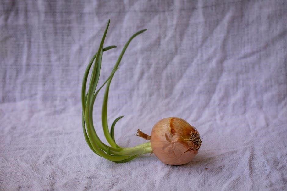
Onion root tips are ideal for studying mitosis due to their rapid cell division‚ large size‚ and accessibility․ The meristematic cells at the root tip divide continuously‚ providing a high concentration of cells in various mitotic stages․ Their transparent cell walls allow for clear visualization under a microscope․ Additionally‚ the simplicity of preparing root tip slides makes them a practical choice for educational labs․ This model system offers insights into the cell cycle‚ making it a cornerstone of biology education for understanding mitosis․

Materials and Equipment Needed for the Lab
Essential materials include onion root tips‚ microscopes‚ glass slides‚ blades‚ and biological stains․ Equipment setup involves preparing slides for cellular observation under magnification․
2․1 List of Required Materials
The materials needed include an onion bulb‚ compound light microscope‚ glass slides‚ cover slips‚ razor blade‚ water‚ iodine or methylene blue stain‚ and a dropper․ Additional items like forceps‚ beakers‚ and paper towels facilitate preparation․ Ensure all equipment is sterilized and ready for use to maintain accuracy and safety during the experiment․
2․2 Equipment Setup and Safety Precautions
Set up the microscope on a stable surface‚ ensuring proper lighting and lens alignment․ Gather all materials‚ including slides‚ stains‚ and tools․ Handle sharp objects like razors with care to avoid cuts․ Wear protective gloves and eyewear when staining․ Ensure good ventilation when using chemicals․ Label slides clearly and dispose of waste properly․ Familiarize yourself with the microscope’s operation before starting․ Follow all lab safety guidelines to minimize risks during the experiment․

Preparation of Onion Root Tip Slide
Begin by selecting a fresh onion root tip‚ then fix and stain the cells to preserve and enhance visibility․ Gently squash the tissue on a slide for microscopic observation․
3․1 Steps for Fixing and Staining the Root Tip
Begin by cutting the onion root tip and immersing it in Carnoy’s fixative to preserve the cells․ After fixation‚ transfer the root tip to a staining solution‚ such as methylene blue or toluidine blue‚ to enhance cell visibility․ Allow the tissue to stain for several minutes before rinsing gently․ This process ensures the cells are properly preserved and prepared for microscopic examination‚ making the mitotic phases easier to identify and study․
3․2 Squashing the Root Tip for Microscopic Observation
Place the stained root tip on a clean slide and add a drop of water․ Using another slide‚ gently squash the root tip to spread the cells evenly․ Apply slight pressure to flatten the tissue‚ ensuring the cells are separated into a single layer․ This step enhances visibility under the microscope by distributing the cells uniformly․ Use forceps to handle the root tip carefully‚ avoiding excessive pressure that could damage the cells․ The goal is to achieve a thin layer of cells for clear microscopic examination․

Observing Mitosis Under the Microscope
Examine the prepared slide under a microscope․ Start with low magnification to locate cells‚ then switch to high magnification for detailed observation of mitotic phases․
4․1 Focusing the Microscope for Clear Visualization
Start by placing the slide under the microscope with the root tip near the center․ Use the coarse focus knob to bring the cells into view at low power․ Once focused‚ switch to high power for clearer details․ Adjust the fine focus for sharp visualization of the cell structures‚ ensuring the mitotic phases are easily distinguishable․ Proper focusing is essential for accurately identifying each stage of mitosis in the onion root tip cells․ This step ensures optimal clarity for detailed observation․
4․2 Identifying Different Phases of Mitosis
Under the microscope‚ identify cells in different mitotic phases․ In interphase‚ the nucleus is visible‚ and chromosomes are uncoiled․ During prophase‚ chromosomes condense‚ and the nuclear envelope breaks down․ In metaphase‚ chromosomes align at the equatorial plate․ Anaphase shows sister chromatids separating toward opposite poles․ Finally‚ in telophase‚ the nuclear envelope reforms‚ and chromosomes uncoil․ Accurately distinguishing these phases is crucial for understanding the mitotic process in onion root tip cells․
Phases of Mitosis in Onion Root Tip Cells
Mitosis in onion root tip cells includes interphase‚ prophase‚ metaphase‚ anaphase‚ and telophase․ Each phase is distinct‚ allowing clear observation of chromosomal changes and cellular division processes․
5․1 Interphase: The Growth and Preparation Phase
Interphase is the longest stage of mitosis‚ where the cell grows‚ replicates its DNA‚ and prepares for cell division․ During this phase‚ the chromatin remains loosely packed‚ allowing transcription․ The cell synthesizes essential proteins and organelles‚ such as centrioles‚ ensuring readiness for division․ This phase is critical for cellular growth and preparation‚ making it a foundational step in the mitotic process․ Observing interphase cells in onion root tips is straightforward due to their large‚ visible nuclei․
5․2 Prophase: Chromosome Condensation and Nuclear Envelope Breakdown
During prophase‚ chromatin condenses into visible chromosomes‚ and a spindle apparatus forms․ The nuclear envelope disintegrates‚ allowing chromosomes to interact with spindle fibers․ This phase prepares the cell for metaphase by organizing genetic material․ In onion root tips‚ prophase cells are identifiable by their condensed chromosomes and the absence of a nuclear membrane‚ making this stage critical for proper cell division and genetic distribution․ Observing prophase highlights the transition from interphase to active mitosis․
5․3 Metaphase: Alignment of Chromosomes at the Equatorial Plate
During metaphase‚ chromosomes align at the equatorial plate‚ attached to spindle fibers․ This ensures equal distribution of genetic material to daughter cells․ The chromosomes are fully condensed and visible under a microscope․ In onion root tips‚ metaphase cells show a clear line of chromosomes across the cell center․ This phase is critical for ensuring genetic continuity and is a key observation point in studying mitosis‚ as it highlights the precise organization of chromosomes before separation․
5․4 Anaphase: Separation of Sister Chromatids
Anaphase marks the separation of sister chromatids‚ pulled to opposite poles by spindle fibers․ This ensures each daughter cell receives identical genetic material․ In onion root tips‚ anaphase cells show chromatids being pulled apart‚ creating two distinct sets of chromosomes; This phase is brief but critical‚ as it ensures genetic continuity and prepares for cytokinesis․ Observing anaphase in root tips highlights the dynamic nature of chromosome movement during mitosis․
5․5 Telophase: Reformation of the Nuclear Envelope
Telophase involves the reformation of the nuclear envelope‚ marking the end of mitosis․ The chromosomes uncoil‚ returning to chromatin form‚ and a nuclear membrane encloses each set of chromosomes․ This phase restores the cell’s nucleus to its interphase state․ In onion root tips‚ telophase cells show the nuclear envelope reassembling‚ ensuring genetic material is organized for the next cell cycle․ This step is crucial for completing cell division and preparing for cytokinesis․

Data Collection and Analysis
This section involves counting cells in each mitotic phase and calculating the time spent in each stage․ It helps analyze the cell cycle duration and its distribution․
6․1 Counting Cells in Each Phase of Mitosis
Count cells in each mitotic phase (interphase‚ prophase‚ metaphase‚ anaphase‚ telophase) using a microscope․ Record the number of cells in each phase to determine the duration spent in each stage․ Use a tally system or table for accurate counting․ Ensure multiple fields of view are observed to gather representative data․ This step helps assess the proportion of cells in each phase‚ providing insights into the cell cycle’s dynamics and the time cells spend in each phase․
6․2 Calculating the Time Spent in Each Phase
Calculate the time spent in each mitotic phase by analyzing cell counts․ Divide the number of cells in each phase by the total number of cells observed to find the percentage․ This percentage reflects the proportion of time cells spend in each phase․ Since mitosis is continuous‚ cells are more likely to be in longer phases like interphase․ This step helps estimate the duration of each phase and provides insights into the cell cycle’s timing and dynamics․

Results and Discussion
The results revealed cells in various mitotic phases‚ with most in interphase․ Observations supported the expected timeline of the cell cycle‚ confirming mitosis dynamics in onion root tips․

7․1 Presentation of Findings
During the mitosis lab‚ most onion root tip cells were found in interphase‚ while fewer cells were observed in the other phases․ The distribution of cells across the phases was not equal‚ with a higher concentration in interphase and a smaller percentage in prophase‚ metaphase‚ anaphase‚ and telophase․ The findings were recorded and presented in tables and charts‚ providing a clear visual representation of the data collected during the experiment․
7․2 Interpretation of Results and Comparison with Expected Outcomes
The results align with expected outcomes‚ as most cells were in interphase‚ while fewer were in prophase‚ metaphase‚ anaphase‚ and telophase․ This reflects the cell cycle’s nature‚ where interphase is the longest phase․ The distribution of cells across phases supports the theory that mitosis is a rapid process compared to interphase․ These findings validate the use of onion root tips for studying mitosis‚ as they provide a clear representation of cell division stages․
The onion root tip mitosis lab effectively demonstrates the stages of cell division‚ providing insights into the cell cycle and its significance in growth and development․
8․1 Summary of Key Observations
The onion root tip mitosis lab revealed cells in various stages of mitosis․ Interphase was most common‚ while prophase‚ metaphase‚ anaphase‚ and telophase were less frequent․ Observations confirmed the presence of chromosomal condensation‚ nuclear envelope breakdown‚ chromosome alignment‚ sister chromatid separation‚ and nuclear reformation․ The distribution of cells across mitotic phases provided insights into the duration of each stage‚ with interphase being the longest․ These findings align with the cell cycle’s characteristics‚ emphasizing the importance of mitosis in plant growth and development․
8․2 Significance of the Experiment in Understanding Cell Division
This experiment is crucial for understanding cell division‚ as it provides a visible demonstration of mitosis․ Observing onion root tips allows students to identify and study the stages of mitosis‚ reinforcing concepts of the cell cycle․ By analyzing the distribution of cells in each phase‚ the experiment highlights the dynamic nature of cellular processes․ Such studies are fundamental in biology‚ offering insights into growth‚ tissue repair‚ and the mechanisms underlying uncontrolled cell division‚ such as cancer․

Additional Resources and Answer Key
This section provides additional resources‚ including lab manuals and guides‚ to deepen understanding of mitosis․ It also includes an answer key for the lab report․
9․1 Links to Lab Manuals and Guides
Several online resources provide detailed lab manuals and guides for conducting the onion root tip mitosis experiment․ Websites like Mrs․ McGaffin’s Biology Resources and educational platforms offer step-by-step instructions․ Additionally‚ PDF documents such as “Experiment 1․1 Mitosis in Onion Root Tip” and “Cell Division: Mitosis in Onion Root Tips” are widely available․ These resources include procedures for slide preparation‚ staining‚ and microscopic observation‚ ensuring a comprehensive understanding of the lab process and its expected outcomes․
9․2 PDF Answer Key for Pre- and Post-Lab Questions
PDF answer keys for pre- and post-lab questions on the onion root tip mitosis lab are available online․ These documents provide correct answers to questions about mitosis phases‚ root tip cell division‚ and lab procedures․ Resources like “Experiment 1․1 Mitosis in Onion Root Tip” and “Cell Division: Mitosis in Onion Root Tips” include detailed answer keys․ These PDFs are essential for students to verify their understanding and prepare for assessments related to the lab experiment․


Be the first to reply